

What Do Proteins Look Like Protein Digestion In The Stomach Animated Gif
Macronutrient Digestion
You probably do not retrieve too much nigh what actually happens to the nutrient you eat. This section will draw in depth how what you consume is digested. The desired end consequence for the learner will be an integrated agreement of the process. This will crave college levels of thinking, but will prove to be well worth it in the end.
Sections:
3.1 Digestion at a Glance
3.ii Mouth to the Tummy
three.three Stomach
3.4 Pocket-size Intestine
3.5 Macronutrient Digestion Review
3.6 Large Intestine
No References
3.one Digestion at a Glance
Digestion is the process of breaking down food to be captivated or excreted. The gastrointestinal (GI, digestive) tract, the passage through which our food travels, is a "tube within a tube." The trunk of our body is the outer tube and the GI tract is the interior tube, as shown beneath. Thus, even though the GI tract is inside the torso, the actual interior of the tract is technically outside of the body. This is considering the contents have to be captivated into the trunk. If it's not absorbed, it will exist excreted and never enter the body itself.
A number of organs are involved in digestion, which collectively are referred to as the digestive system.
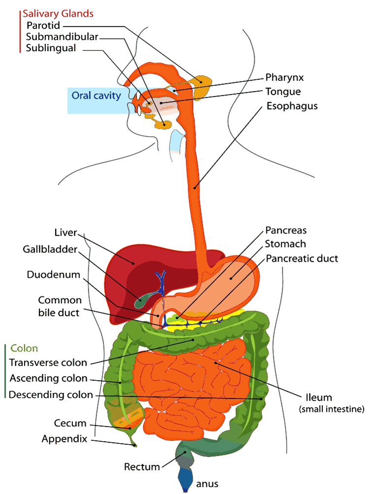
The organs that grade the alimentary canal (mouth, esophagus, stomach, small intestine, big intestine (aka colon), rectum, and anus) come into direct contact with the nutrient or digestive content.
The journey through the gastrointestinal tract starts in the mouth and ends in the anus as shown beneath:
Mouth –> Esophagus –> Stomach –> Small Intestine –> Big Intestine –> Rectum –> Anus
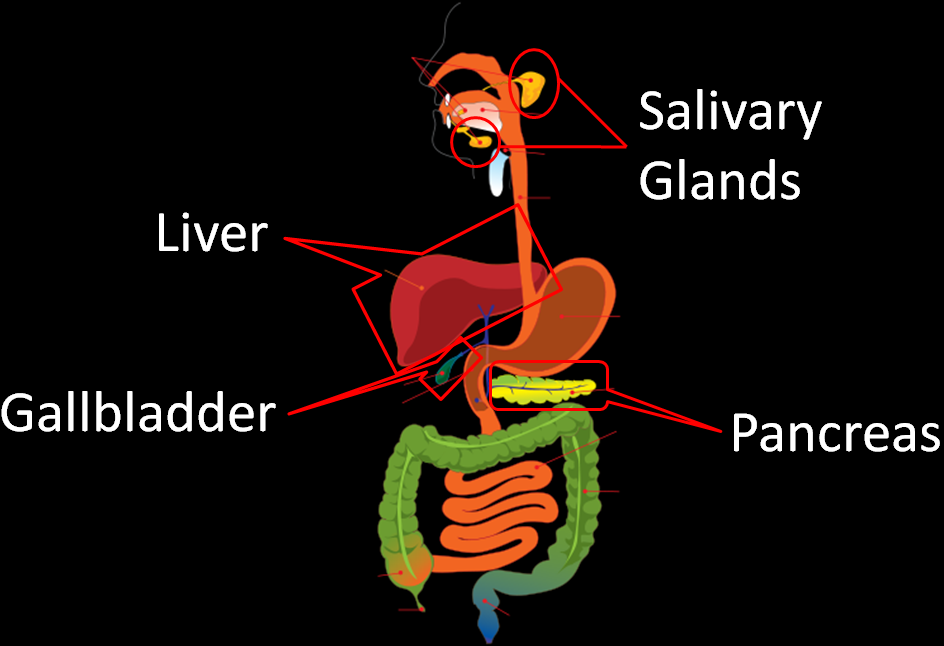
In addition to the GI tract, there are digestion accompaniment organs (salivary glands, pancreas, gallbladder, and liver) that play an integral role in digestion. The accessory organs exercise non come directly in contact with nutrient or digestive content.
At that place are a number of enzymes that are involved in digestion. We will get through each i in detail, but this table should assist give an overview of which enzymes are active at each location of the GI tract.
Table 3.11 Digestive enzymes
| Location | Enzyme/Coenzyme |
| Mouth | Salivary amylase Lingual lipase |
| Stomach | Pepsin Gastric lipase |
| Small-scale Intestine | Pancreatic alpha-amylase Brush border disaccharidases Pancreatic lipase Colipase Phospholipase-A2 Cholesterol esterase Proteases Brush edge peptidases |
References & Links
- http://www.wpclipart.com/medical/anatomy/digestive/Digestive_system_diagram_page.png.html
- http://eatables.wikimedia.org/wiki/File:Digestivetract.gif
Video
Enzymes and Digestion – http://www.youtube.com/watch?v=bNMsNHqxszc
3.two Mouth to the Stomach
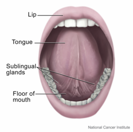
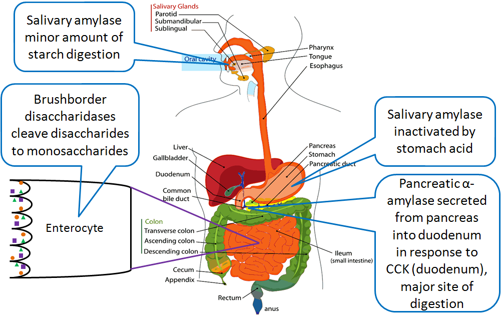
Digestion begins in the oral cavity, both mechanically and chemically. Mechanical digestion is called mastication, or the chewing and grinding of food into smaller pieces. The salivary glands release saliva, mucus, and the enzymes, salivary amylase and lysozyme.
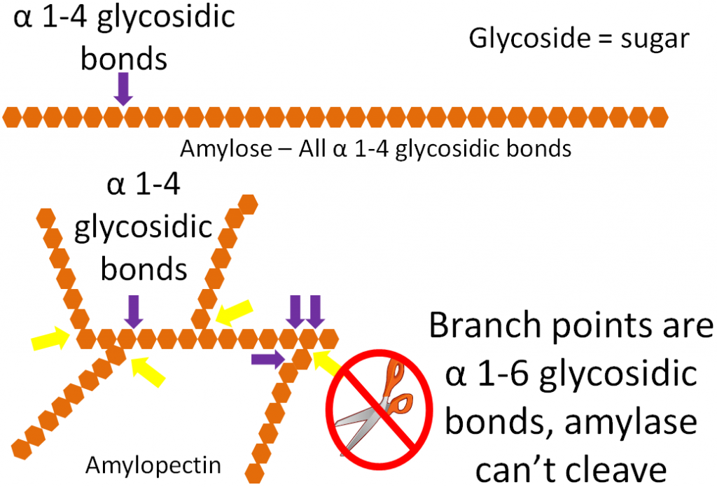
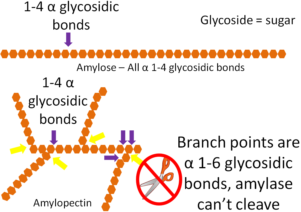
Salivary amylase cleaves the blastoff i-4 glycosidic bonds in the starch molecules, amylose and amylopectin. Notwithstanding, salivary amylase cannot cleave the branch points in amylopectin where there are alpha 1-vi glycosidic bonds, as shown in the figure beneath. Overall this enzyme accounts for a minor amount of carbohydrate digestion.
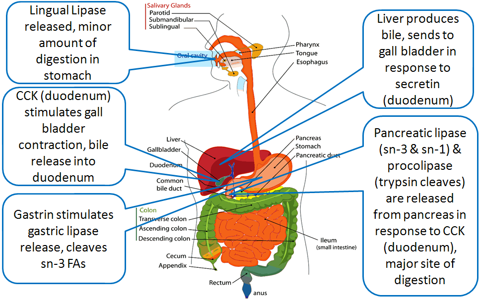
Lysozyme helps suspension downwards leaner prison cell walls to prevent a possible infection. Another enzyme, lingual lipase, is also released in the mouth. Although it is released in the mouth, it is most agile in the stomach where it preferentially cleaves short-chain fat acids in the sn-3 position. Lingual lipase has a small role in digestion in adults, just may be important for infants to assist pause down triglycerides in chest milk2.
Swallowing
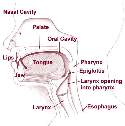
Now that the food has been thoroughly chewed and formed into a bolus, it can proceed downwards the throat to the side by side stop in digestion. It will motility down the pharynx where it reaches a "fork in the route", with the larynx as one route and the esophagus every bit the other. The esophagus road leads to the stomach; this is the management that nutrient should go. The other road, through the larynx, leads to the trachea and ultimately the lungs. This is definitely not where you want your nutrient or drink going, as this is the pathway for the air y'all breathe.
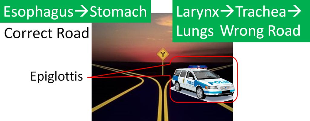
Fortunately, our body was designed in such a way that a small tissue, called the epiglottis, covers the opening to the trachea. It directs the food downward the correct route equally shown beneath.
Esophagus
Before beingness correctly guided into the esophagus, the bolus of food will travel through the upper esophageal sphincter. Sphincters are circular muscles that are constitute throughout the gastrointestinal tract that essentially serve as gates between the different sections. Once in the esophagus, wavelike muscular movements, known as peristalsis, occur, as shown in the animation and video in the links beneath.
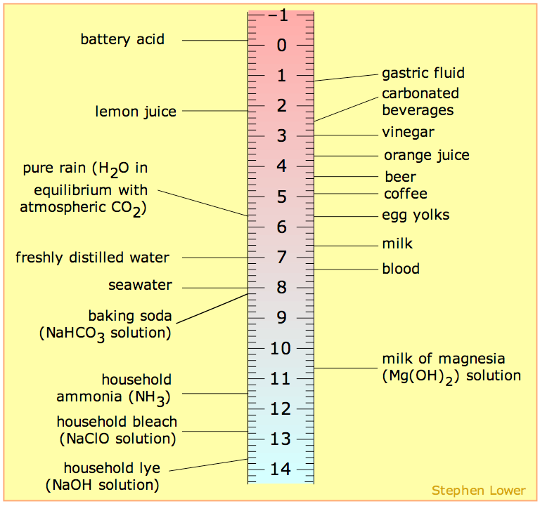
At the end of the esophagus the bolus volition run across the lower esophageal sphincter. This sphincter keeps the harmful acids of the stomach out of the esophagus. However, in many people this sphincter is leaky, which allows stomach acid to reflux, or creep upward, the esophagus. Stomach acid is very acidic (has a low pH). The ruler below will give you an thought of just how acidic the breadbasket is. Notice that the pH of gastric (term used to describe the breadbasket) fluid is lower (more acidic) than any of the listed items as well battery acrid.

The leaking of the very acidic gastric contents results in a burning awareness, commonly referred to as "heartburn." If this occurs more than twice per week and is severe, the person may have gastroesophageal reflux disease (GERD). The following videos explain more virtually these conditions.
Tabular array 3.21 Review of Chemical Digestion in the Mouth
| Macronutrient | Action |
| Carbohydrates | Salivary amylase cleaves ane,4-glycosidic bonds |
| Lipids | Release of lingual lipase |
| Protein | None |
References & Links
- Alan Hoofring, http://visualsonline.cancer.gov/details.cfm?imageid=4371
- Shils ME, Shike Grand, Ross Air conditioning, Caballero B, Cousins RJ, editors. (2006) Modern diet in health and disease. Baltimore, MD: Lippincott Williams & Wilkins.
- http://en.wikipedia.org/wiki/File:Illu01_head_neck.jpg
- http://upload.wikimedia.org/wikipedia/eatables/4/46/PH_scale.png
Link
Peristalsis – http://en.wikipedia.org/wiki/File:Peristalsis.gif
Videos
Peristalsis Animation – http://www.youtube.com/watch?5=o18UycWRsaA
Acrid Reflux – https://www.youtube.com/sentry?five=SW-QfyDSY5I
GERD 101 – http://www.youtube.com/watch?v=FqdOvZkrSYk&feature=rec-lis-watch-cur_emp-farside_rn
3.3 Breadbasket
Afterward going through the lower esophageal sphincter, food enters the stomach. Our stomach is involved in both chemical and mechanical digestion. Mechanical digestion occurs as the stomach churns and grinds nutrient into a semisolid substance called chyme (partially digested food).
The lining of the tummy is made upward of dissimilar layers of tissue. The mucosa is the outermost layer (closest to stomach crenel) as shown in the figure beneath.
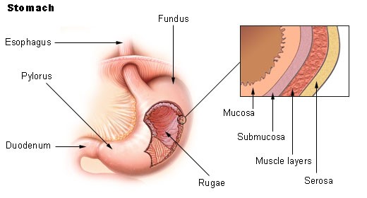
The mucosa is not a flat surface. Instead, its surface is lined by gastric pits, as shown in the effigy beneath.
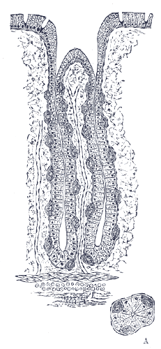
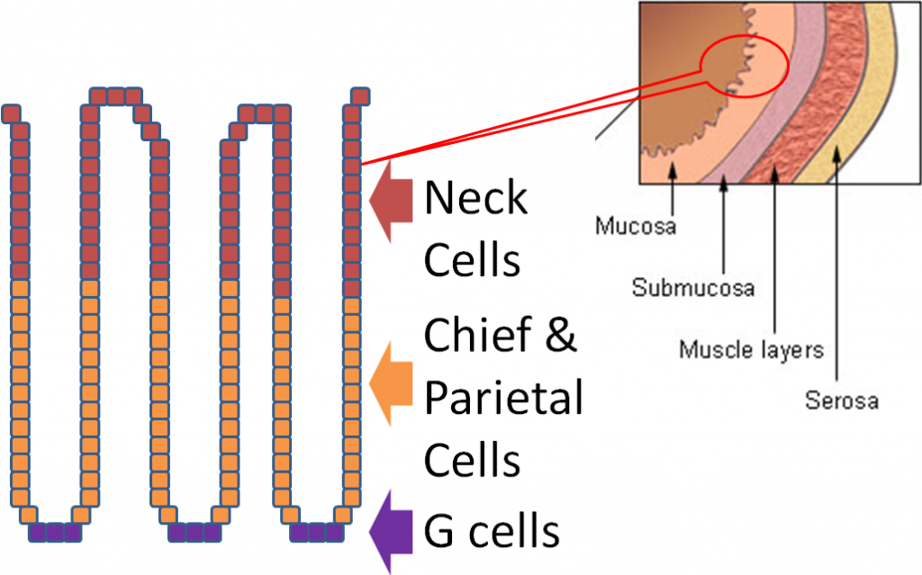
Gastric pits are indentations in the tum's surface that are lined past 4 different types of cells.
The following video is a nice introduction to gastric pits and talks near chief and parietal cells that are covered in more detail below.
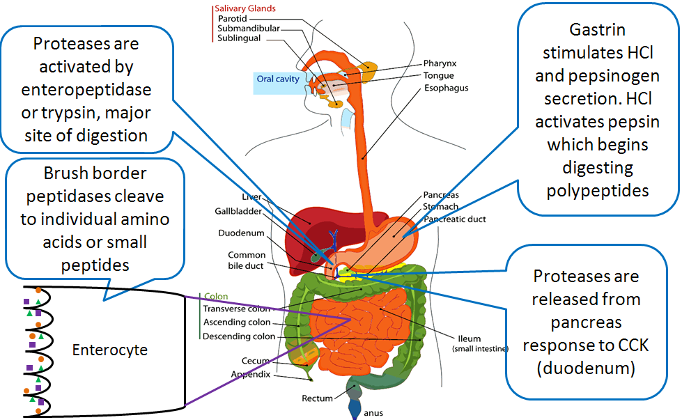
At the bottom of the gastric pit are the G cells that secrete the hormone gastrin. Gastrin stimulates the parietal and chief cells that are found above the 1000 cells. The chief cells secrete the zymogen pepsinogen and the enzyme gastric lipase. A zymogen is an inactive forerunner of an enzyme that must be cleaved or contradistinct to form the active enzyme. The parietal cells secrete hydrochloric acid (HCl), which lowers the pH of the gastric juice (water + enzymes + acrid). The HCl inactivates salivary amylase and catalyzes the conversion of pepsinogen to pepsin. Finally, the tiptop of the pits are the neck cells that secrete mucus to prevent the gastric juice from digesting or dissentious the stomach mucosa3. The table below summarizes the actions of the different cells in the gastric pits.
Tabular array 3.41 Cells involved in the digestive processes in the stomach
| Blazon of Cell | Secrete |
| Cervix | Mucus |
| Chief | Pepsinogen and gastric lipase |
| Parietal | HCl |
| G | Gastrin |
The figure below shows the action of all these different secretions in the stomach.
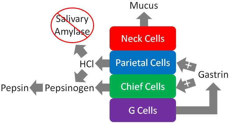
To reiterate, the figure to a higher place illustrates that the cervix cells of the gastric pits secrete mucus to protect the mucosa of the stomach from essentially digesting itself. Gastrin from the Grand cells stimulates the parietal and chief cells to secrete HCl and enzymes, respectively.
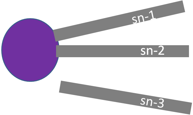
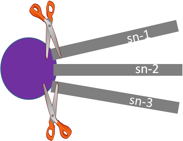
The HCl in the stomach denatures salivary amylase and other proteins by breaking down the structure and, thus, the function of it. HCl as well converts pepsinogen to the active enzyme pepsin. Pepsin is a protease, significant that it cleaves bonds in proteins. Information technology breaks down the proteins in food into individual peptides (shorter segments of amino acids). The other enzyme that is active in the tum is gastric lipase. This enzyme preferentially cleaves the sn-3 position of triglycerides to produce one,2-diglyceride and a free fatty acrid, every bit shown below4. It is responsible for upward to twenty% of triglyceride digestion3.
The chyme volition and so get out the stomach and enter the small intestine via the pyloric sphincter (shown below).
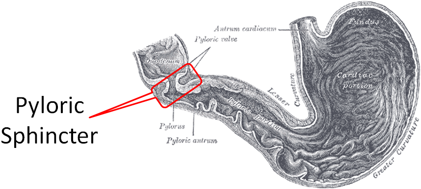
Table 3.32 Summary of chemical digestion in the stomach
| Chemical or Enzyme | Activeness |
| Gastrin | Stimulates chief cells to release pepsinogen Stimulates parietal cells to release HCl |
| HCl | Denatures salivary amylase Denatures proteins Activates pepsinogen to pepsin |
| Pepsin | Cleaves proteins to peptides |
| Gastric lipase | Cleaves sn-iii FA of triglycerides |
References & Links
- https://en.wikipedia.org/wiki/Stomach#/media/File:Illu_stomach2.jpg
- http://en.wikipedia.org/wiki/File:Gray1055.png
- Gropper SS, Smith JL, Groff JL. (2008) Advanced nutrition and human metabolism. Belmont, CA: Wadsworth Publishing.
- Stipanuk MH. (2006) Biochemical, physiological, & molecular aspects of human nutrition. St. Louis, MO: Saunders Elsevier.
- https://en.wikipedia.org/wiki/Pylorus#/media/File:Gray1050.png
Video
Gastric Pits – http://www.youtube.com/watch?v=6hquzCXYlNg
three.4 Small Intestine
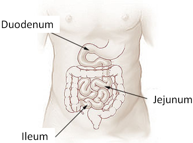
The small intestine is the primary site of digestion. It is divided into 3 sections: the duodenum, jejunum, and ileum (shown below). After leaving the stomach, the outset function of the small intestine that chyme will encounter is the duodenum.
The small intestine consists of many layers, which can be seen in the cross section below.
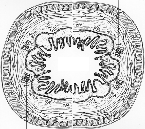
Examining these layers closer, we are going to focus on the epithelium, which comes into contact with the chyme and is responsible for assimilation. The lumen is the name of the cavity that is considered "outside the torso" that chyme moves through.
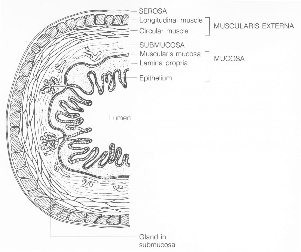
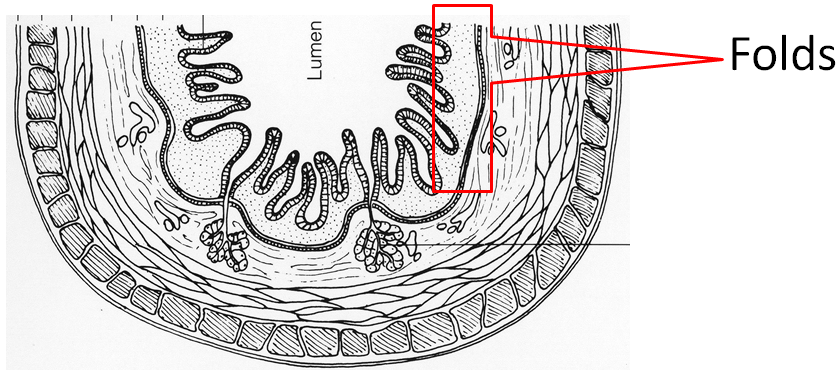
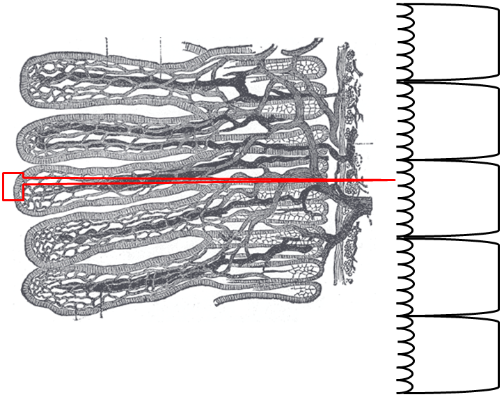
The organization of the minor intestine is in such a way that information technology contains circular folds and finger-like projections known as villi. The folds and villi are shown in the next few figures.
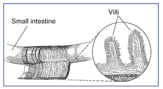
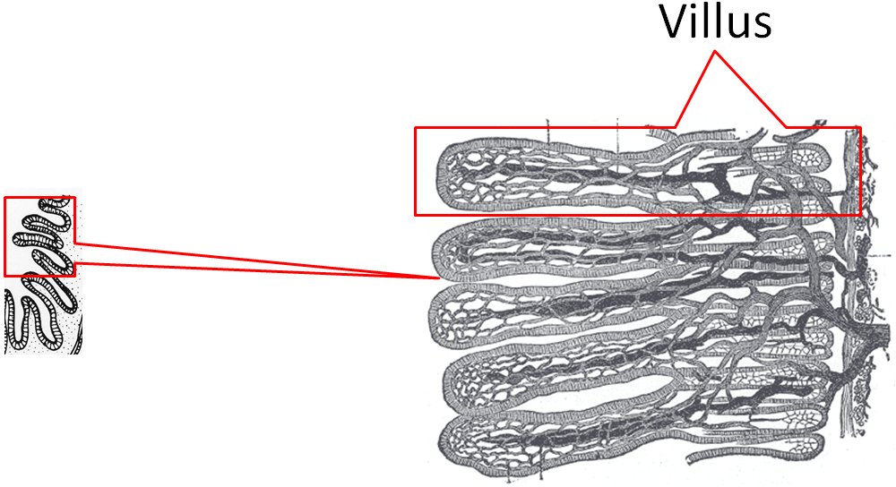
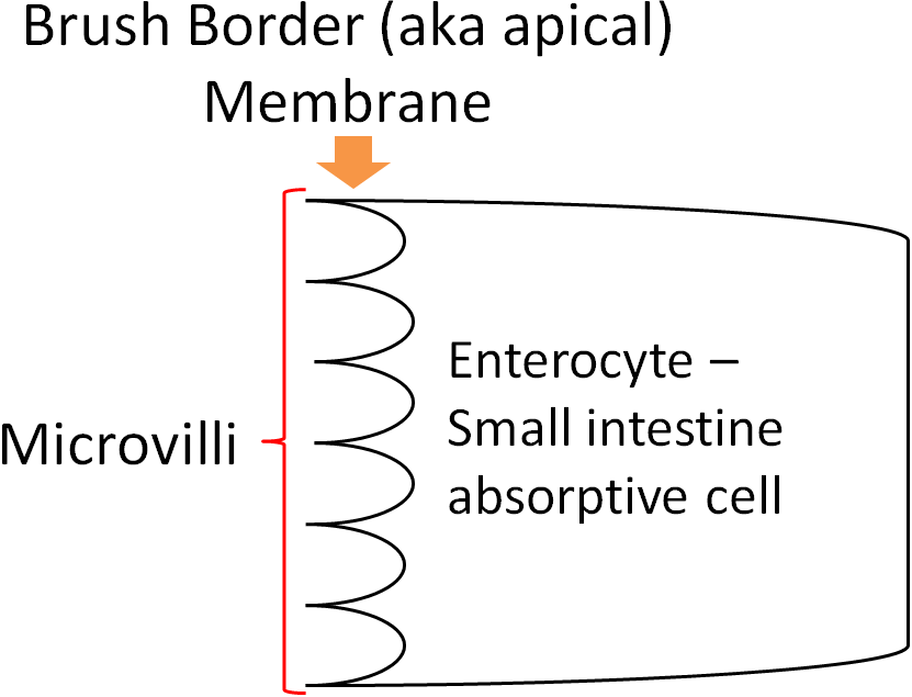
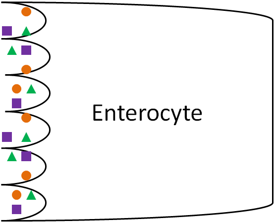
If we were to zoom in even closer, nosotros would be able to see that enterocytes (small intestine absorptive cells) line villi equally shown below.
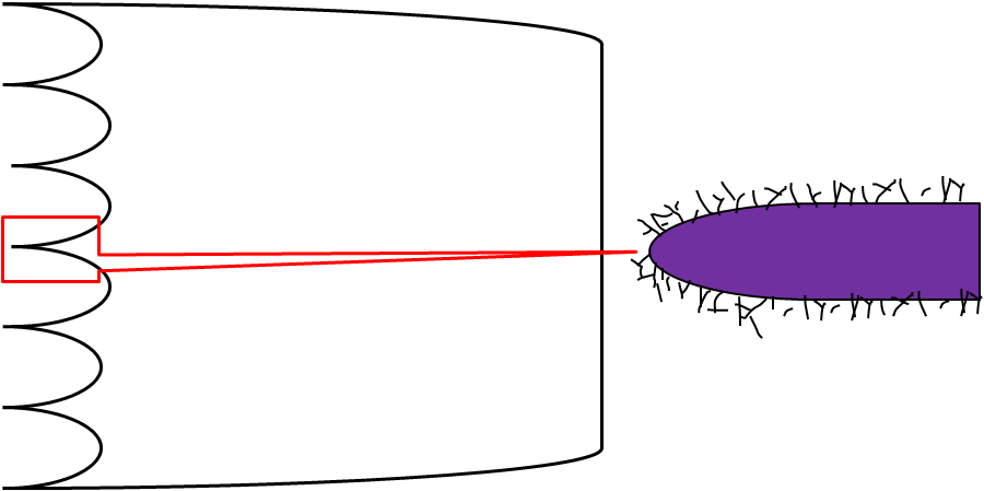
The side, or membrane, of the enterocyte that faces the lumen is not shine either. It is lined with microvilli, and is known as the castor border (aka apical) membrane, as shown below.
Together these features (folds + villi + microvilli) increase the surface surface area ~600 times versus if it was a polish tube5. More than surface area leads to more than contact with the enterocytes and thus, increased absorption.
Going even closer, we discover that the surface of the microvilli is covered by the hair-similar glycocalyx, which is glycoproteins and carbohydrates as shown below.
Now that you take learned about the anatomy of the small intestine, the post-obit subsections get through the different digestive processes that occur at that place.
Subsections:
iii.41 Digestive Hormones, Accessory Organs, & Secretions
three.42 Saccharide Digestion in the Small Intestine
3.43 Poly peptide Digestion in the Small-scale Intestine
iii.44 Lipid Digestion in the Pocket-size Intestine
References & Links
- http://commons.wikimedia.org/wiki/Prototype:Illu_small_intestine_catal%C3%A0.png
- Writer unknown, NCI, http://visualsonline.cancer.gov/details.cfm?imageid=1781
- http://digestive.niddk.nih.gov/ddiseases/pubs/celiac/
- http://commons.wikimedia.org/wiki/Image:Gray1061.png
- Byrd-Bredbenner C, Moe G, Beshgetoor D, Berning J. (2009) Wardlaw's perspectives in nutrition. New York, NY: McGraw-Hill.
three.41 Digestive Hormones, Accessory Organs & Secretions
Earlier we become into the digestive details of the minor intestine, it is of import that y'all have a basic understanding of the anatomy and physiology of the post-obit digestion accessory organs: pancreas, liver, and gallbladder. Digestion accessory organs assist in digestion, just are not office of the gastrointestinal tract. How are these organs involved?
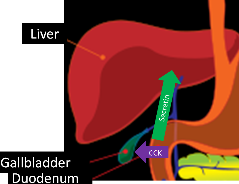
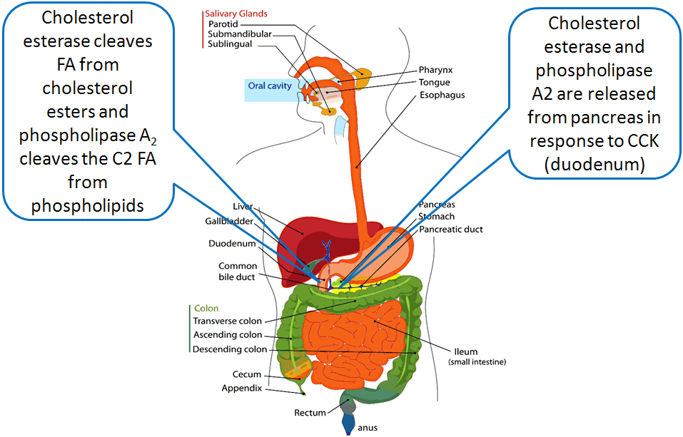
Upon inbound the duodenum, the chyme causes the release of ii hormones from the modest intestine: secretin and cholecystokinin (CCK, previously known every bit pancreozymin) in response to acid and fat, respectively. These hormones have multiple furnishings on different tissues. In the pancreas, secretin stimulates the secretion of bicarbonate (HCO3), while CCK stimulates the secretion of digestive enzymes. The bicarbonate and digestive enzymes released together are collectively known every bit pancreatic juice, which travels to the small intestine, every bit shown below.
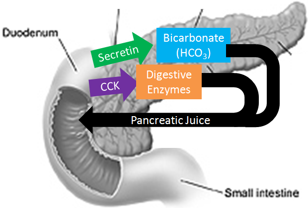
In addition, CCK also stimulates the contraction of the gallbladder causing the secretion of bile into the duodenum.
Pancreas
The pancreas is found behind the stomach and has ii different portions. Information technology has an endocrine (hormone-producing) portion that contains alpha and beta cells that secrete the hormones glucagon and insulin, respectively. However, the vast majority of the pancreas is made up of acini, or acinar cells, that are responsible for producing pancreatic juice. The following video does a nice chore of showing and explaining the role of the different pancreatic cells.
Bicarbonate is a base of operations (high pH) meaning that it can aid neutralize acid. You can discover sodium bicarbonate (NaHCO3, baking soda) on the ruler below to get an idea of its pH.
The chief digestive enzymes in pancreatic juice are listed in the table below. Their function volition be discussed further in subsequently subsections.
Table 3.411 Enzymes in pancreatic juice
| Enzyme |
| Pancreatic alpha-amylase |
| Proteases |
| Pancreatic Lipase & Procolipase* |
| Phospholipase A2 |
| Cholesterol Esterase |
*Not an enzyme
Liver
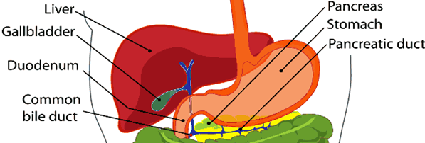
The liver is the largest internal and nigh metabolically active organ in the body. The figure below shows the liver and the accompaniment organs position relative to the stomach.
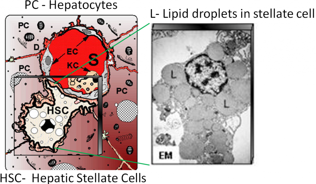
The liver is fabricated up two major types of cells. The principal liver cells are hepatocytes, which conduct out most of the liver'south functions. Hepatic is another term for liver. For example, if yous are going to refer to liver concentrations of a certain food, these are often reported equally hepatic concentrations. The other major jail cell type is the hepatic stellate (also known as Ito) cells. These are fat storing cells in the liver. These ii cell types are depicted below.
The liver'southward major role in digestion is to produce bile. This is a dark-green-yellow fluid that is composed primarily of bile acids, merely also contains cholesterol, phospholipids, and the pigments bilirubin and biliverdin. Bile acids are synthesized from cholesterol. The 2 primary bile acids are chenodeoxycholic acid and cholic acid. In the same way that fatty acids are found in the form of salts, these bile acids tin also be found equally salts. These salts have an (-ate) ending, as shown below.
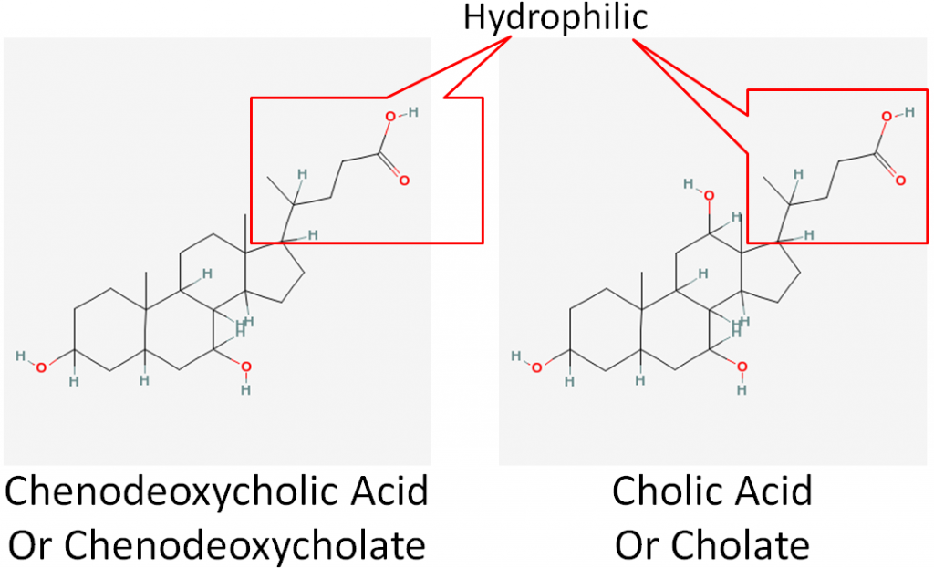
Bile acids, much similar phospholipids, have a hydrophobic and hydrophilic end. This makes them splendid emulsifiers that are instrumental in fat digestion. Bile is and so transported to the gallbladder.
Gallbladder
The gallbladder is a small, sac-like organ found just off the liver (see figures to a higher place). Its primary function is to shop and concentrate bile fabricated by the liver. The bile is then transported to the duodenum through the mutual bile duct.
Why practice nosotros need bile?
Bile is important considering fat is hydrophobic and the surroundings in the lumen of the pocket-size intestine is watery. In add-on, in that location is an unstirred water layer that fat must cross to reach the enterocytes in social club to be absorbed.
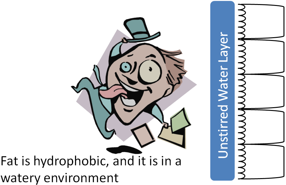
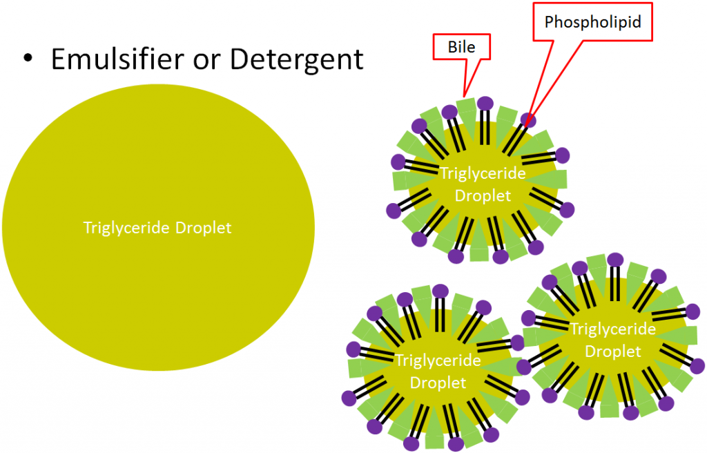
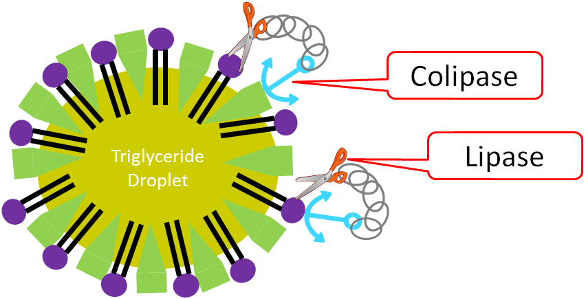
Here triglycerides form large triglyceride droplets to go on the interaction with the watery environment to a minimum. This is inefficient for digestion, considering enzymes cannot admission the interior of the droplet. Bile acts as an emulsifier, or detergent. It, forth with phospholipids, forms smaller triglyceride droplets that increase the surface area that is attainable for triglyceride digestion enzymes, as shown below.
Secretin and CCK also command the production and secretion of bile. Secretin stimulates the flow of bile from the liver to the gallbladder. CCK stimulates the gallbladder to contract, causing bile to be secreted into the duodenum, as shown below.
References & Links
- Don Bliss, NCI, http://visualsonline.cancer.gov
- http://upload.wikimedia.org/wikipedia/eatables/4/46/PH_scale.png
- http://www.wpclipart.com/medical/anatomy/digestive/Digestive_system_diagram_page.png.html
- http://www.comparative-hepatology.com/content/half dozen/ane/7
Video
The Pancreas – http://world wide web.youtube.com/watch?v=j5WF8wUFNkI
3.42 Sugar Digestion in the Small Intestine
The small intestine is the primary site of carbohydrate digestion. Pancreatic blastoff-amylase is the primary carbohydrate digesting enzyme. Pancreatic alpha-amylase, like salivary amylase, cleaves the alpha i-iv glycosidic bonds of carbohydrates, reducing them to simpler carbohydrates, such as glucose, maltose, maltotriose, and dextrins (oligosaccharides containing ane or more blastoff one-6 glycosidic bonds). Pancreatic amylase is also unable to carve the branch point alpha ane-half dozen bonds1.
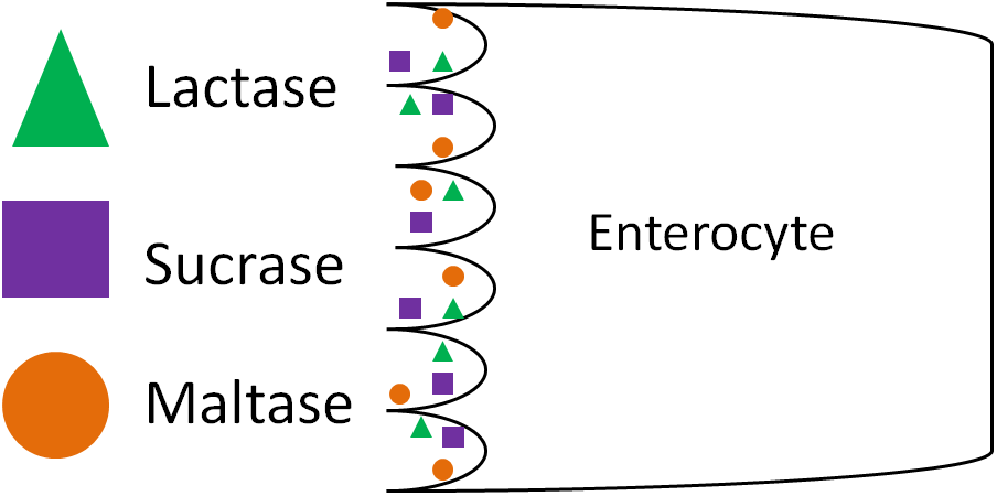
The pancreatic amylase products, along with the disaccharides sucrose and lactose, and so move to the surface of the enterocyte. Hither, there are disaccharidase enzymes (lactase, sucrase, maltase) on the outside of the enterocyte. Enzymes, like these, that are on the outside of cell walls are referred to every bit ectoenzymes. Individual monosaccharides are formed when lactase cleaves lactose, sucrase cleaves sucrose, and maltase cleaves maltose. In that location is also another castor border enzyme, alpha-dextrinase. This enzyme cleaves blastoff 1-6 glycosidic bonds in dextrins, primarily the co-operative point bonds in amylopectin. The products from these brush edge enzymes are the unmarried monosaccharides glucose, fructose, and galactose that are ready for absorption into the enterocyte1.
References & Links
- Gropper SS, Smith JL, Groff JL. (2008) Advanced nutrition and homo metabolism. Belmont, CA: Wadsworth Publishing.
3.43 Protein Digestion in the Small Intestine
The small intestine is the major site of protein digestion by proteases (enzymes that carve proteins). The pancreas secretes a number of proteases as zymogens into the duodenum where they must be activated earlier they tin carve peptide bonds1. This activation occurs through an activation cascade. A cascade is a series of reactions in which one step activates the side by side in a sequence that results in an distension of the response. An example of a cascade is shown beneath.
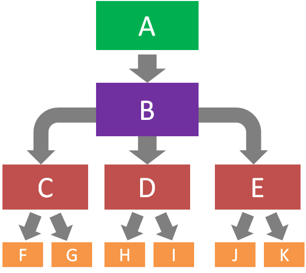
In this example, A activates B, B activates C, D, and Due east, C activates F and One thousand, D activates H and I, and E activates G and 50. Cascades also help to serve as control points for certain process. In the protease cascade, the activation of B is really important because it starts the cascade.
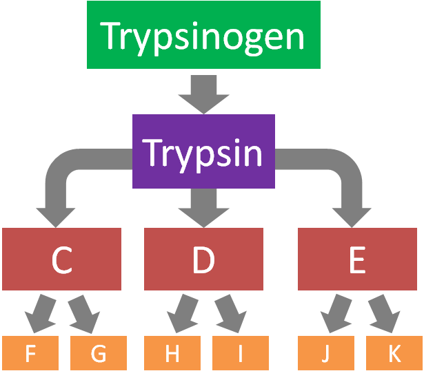
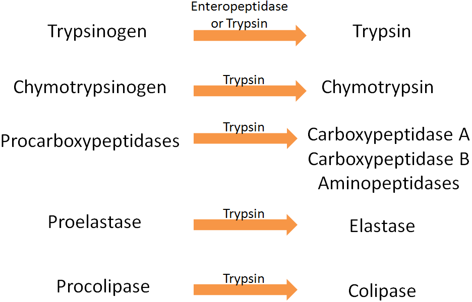
The protease/colipase activation scheme starts with the enzyme enteropeptidase (secreted from the intestinal brush border) that converts trypsinogen to trypsin. Trypsin can activate all the proteases (including itself) and colipase (involved in fatty digestion)1 every bit shown in the 2 figures below.
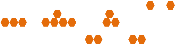
The products of the activeness of the proteases on proteins are dipeptides, tripeptides, and individual amino acids, every bit shown beneath.
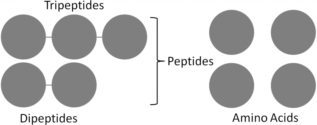
At the brush border, much like disaccharidases, there are peptidases that cleave some peptides down to amino acids. Not all peptides are cleaved to private amino acrid, because small peptides tin be taken up into the enterocyte, thus, the peptides practice not need to be completely broken down to individual amino acids. Thus the end products of protein digestion are primarily dipeptides and tripeptides, along with individual amino acids1.
References & Links
- Gropper SS, Smith JL, Groff JL. (2008) Advanced diet and human metabolism. Belmont, CA: Wadsworth Publishing.
iii.44 Lipid Digestion in the Small Intestine
The small intestine is the major site for lipid digestion. There are specific enzymes for the digestion of triglycerides, phospholipids, and cleavage of esters from cholesterol. Nosotros will look at each in this section.
Triglycerides
The pancreas secretes pancreatic lipase into the duodenum as office of pancreatic juice. This major triglyceride digestion enzyme preferentially cleaves the sn-1 and sn-three fatty acids from triglycerides. This cleavage results in the formation of a 2-monoglyceride and 2 free fatty acids as shown beneath.
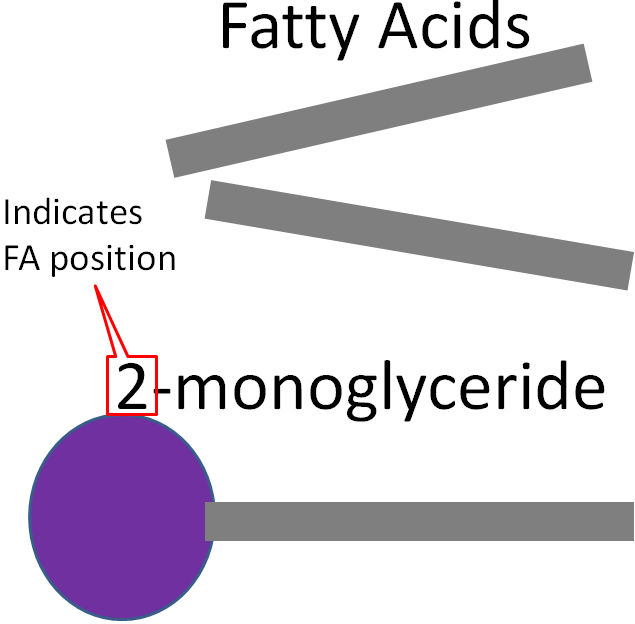
To aid lipase, colipase serves equally an anchor point to help lipase attach to the triglyceride droplet.
Phospholipids
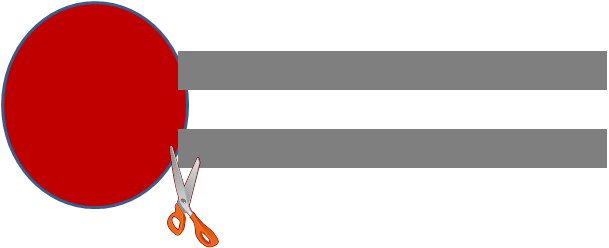
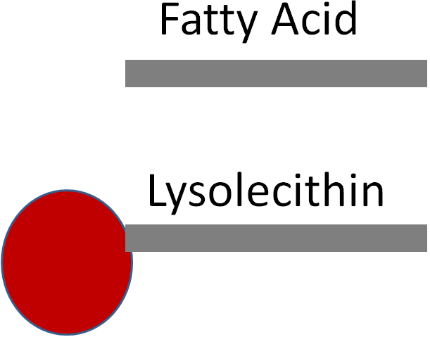
The enzyme phospholipase A2 cleaves the C-ii fatty acid of lecithin, producing lysolecithin and a complimentary fatty acid.
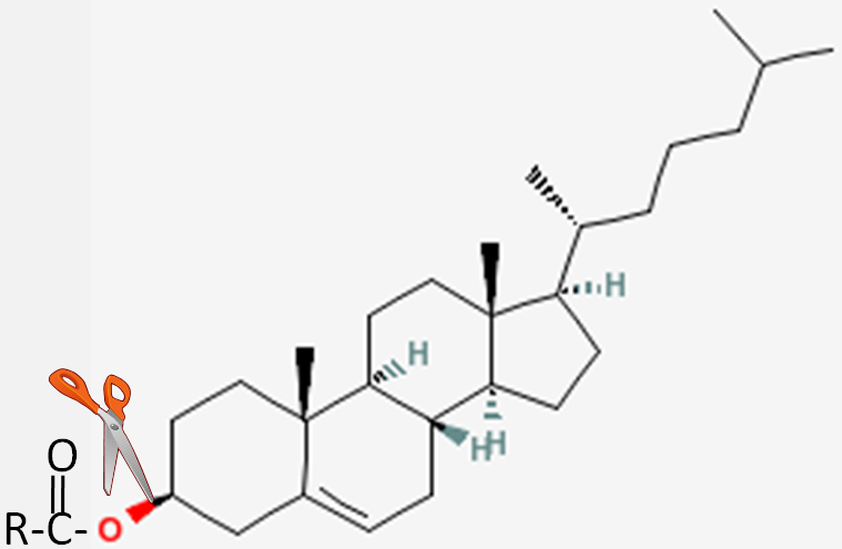
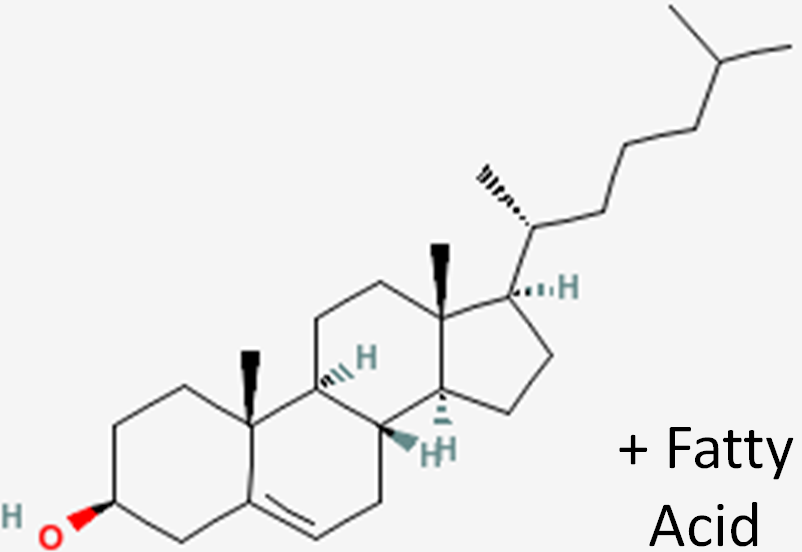
Cholesterol Esters
The fatty acid in cholesterol esters is broken past the enzyme, cholesterol esterase, producing cholesterol and a gratis fatty acrid.
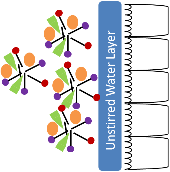
Formation of Mixed Micelles
If zippo else happened at this point, the two-monoglycerides and fatty acids produced by pancreatic lipase would class micelles. The hydrophilic heads would be outward and the fatty acids would exist cached on the interior. These micelles are not sufficiently h2o-soluble to cantankerous the unstirred water layer to get to the brush border of enterocytes. Thus, mixed micelles are formed containing cholesterol, bile acids, and lysolecithin in addition to the 2-monoglycerides and fatty acids, equally illustrated below1.
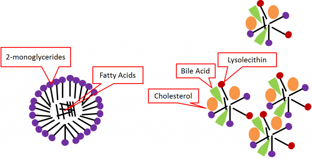
Mixed micelles are more water-soluble, assuasive them to cross the unstirred water layer to the castor border of enterocytes for absorption.
References & Links
- Gropper SS, Smith JL, Groff JL. (2008) Advanced nutrition and human metabolism. Belmont, CA: Wadsworth Publishing.
iii.5 Macronutrient Digestion Review
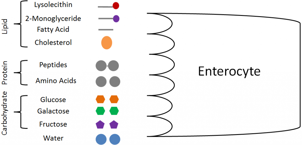
The following figures review the digestion of the unlike macronutrients.
Carbohydrate Digestion
Protein Digestion
Lipid Digestion
Cholesterol Ester and Phospholipid Digestion
After digestion, the products below are ready for uptake into the enterocyte.
References & Links
- http://www.wpclipart.com/medical/anatomy/digestive/Digestive_system_diagram_page.png.html
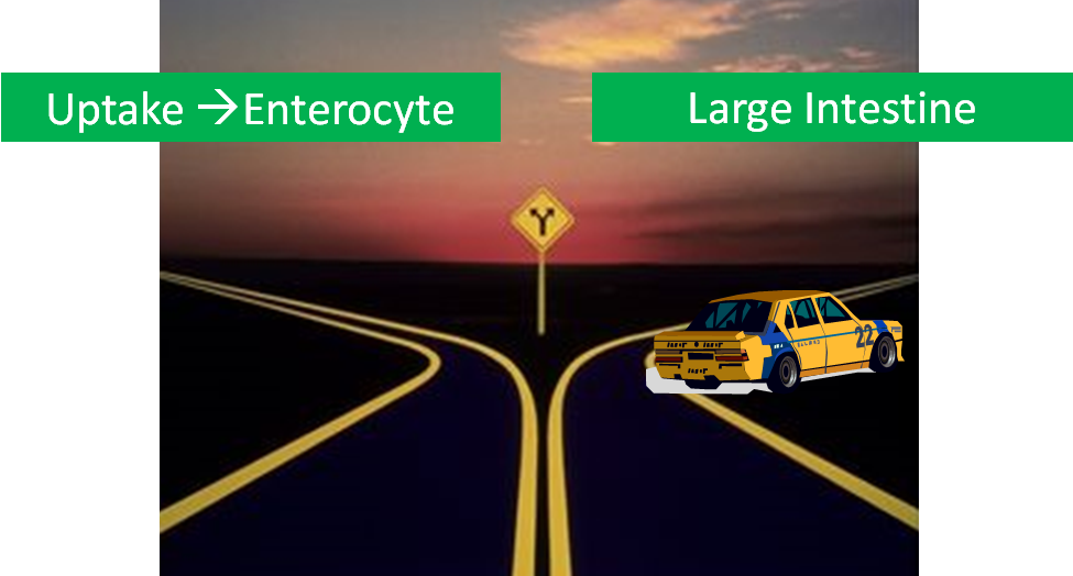
3.6 The Large Intestine
We have reached a fork in the road. We could follow the uptake of the digested compounds into the enterocyte or nosotros could finish post-obit what has escaped digestion and is going to keep into the big intestine. Obviously from the title of this section we are going to do the latter. Equally we learned previously, fiber is a rough term for what has survived digestion and has reached the large intestine.
The ileocecal valve is the sphincter betwixt the ileum and the large intestine. This name should make more than sense as we go through the anatomy of the large intestine.
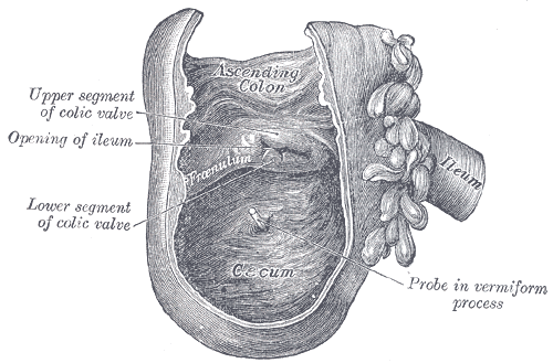
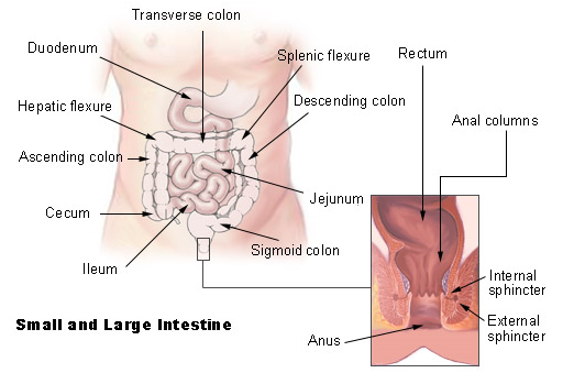
The large intestine consists of the colon, the rectum, and the anus. The colon tin can be further divided into the cecum (hence the -cecal in ileocecal valve, ileo- refers to ileum), ascending colon, transverse colon, descending colon, and sigmoid colon equally shown beneath.
The large intestine is responsible for absorbing remaining water and electrolytes (sodium, potassium, and chloride). It besides forms and excretes feces. The large intestine contains large amounts of microorganisms similar those shown in the figure below.
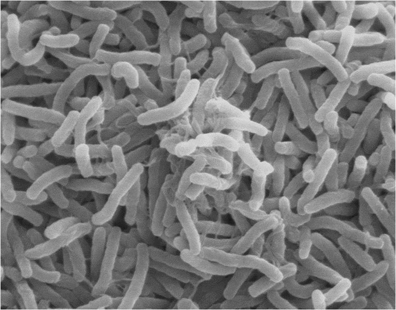
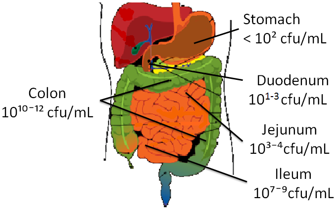
The big intestine can also exist referred to as the gut. There are a large number of microorganisms found throughout the gastrointestinal tract that collectively are referred to equally the flora, microflora, biota, or microbiota. Technically, microbiota is the preferred term because flora ways "pertaining to plants". At that place are 10 times more microorganisms in the gastrointestinal tract than cells in the whole human body4. Every bit can be seen in the figure below, the density of microorganisms increases as you movement downwardly the digestive tract.
As described in the fiber sections, there are two different fates for fiber in one case information technology reaches the large intestine. The fermentable, gluey fiber is fermented past leaner. Fermentation is the metabolism of compounds by the microorganisms in the gut. An example of fermentation is the utilization of the oligosaccharides raffinose and stachyose by microorganisms that results in the product of gas, which can atomic number 82 to flatulence. Too, some bile acids are fermented by microorganisms to form secondary bile acids that can be reabsorbed. These secondary bile acids stand for approximately 20% of the full bile acids in our trunk. Fermentable fibers can be used to class short-chain fat acids that can then be captivated and used by the body. The nonfermentable, nonviscous cobweb is not really altered and will exist a component of carrion, that is and so excreted through the rectum and anus. This process involves both an internal and external sphincter that are shown in figure iii.63 above.
Subsection:
3.61 Probiotics & Prebiotics
References & Links
- https://commons.wikimedia.org/wiki/File:Gray1075.png
- http://en.wikipedia.org/wiki/Image:Illu_intestine.jpg
- http://commons.wikimedia.org/wiki/Prototype:Cholera_bacteria_SEM.jpg
- Guarner F, Malagelada J. (2003) Gut flora in health and illness. The Lancet 361(9356): 512.
- DiBaise J, Zhang H, Crowell Grand, Krajmalnik-Brown R, Decker , et al. (2008) Gut microbiota and its possible relationship with obesity. Mayo Clin Proc 83(4): 460.
- Adapted from: http://www.wpclipart.com/medical/anatomy/digestive/Digestive_system_diagram_page.png.html
iii.61 Probiotics & Prebiotics
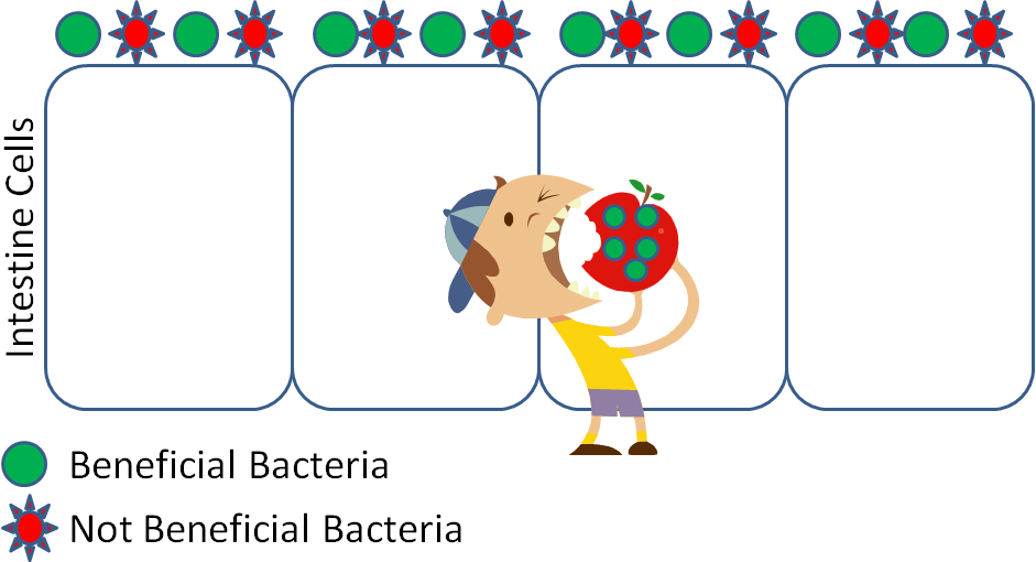
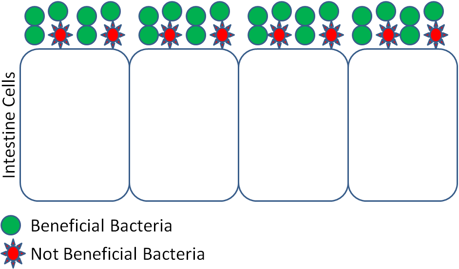
Recently there has been increased attention given to the potential of a person's microbiota to impact health. This is because there are beneficial and non-beneficial bacteria inhabiting our gastrointestinal tracts. Thus, theoretically, if you can increase the benign or decrease the non- beneficial bacteria, in that location may be improved health outcomes. In response to this, probiotics and prebiotics have been identified/developed. A probiotic is a alive microorganism that is consumed, and colonizes in the body as shown in the figures below.
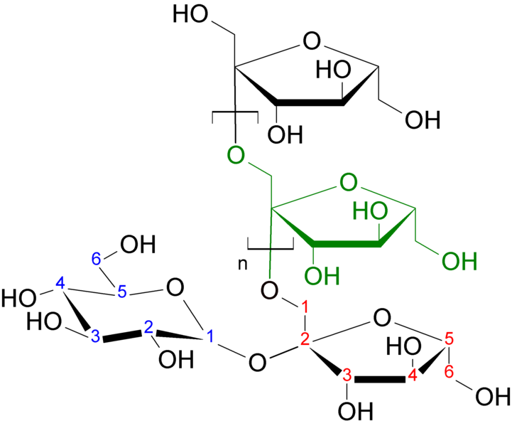
A prebiotic is a nondigestible food component that selectively stimulates the growth of beneficial intestinal bacteria. An example of a prebiotic is inulin, which is shown in the figure below.
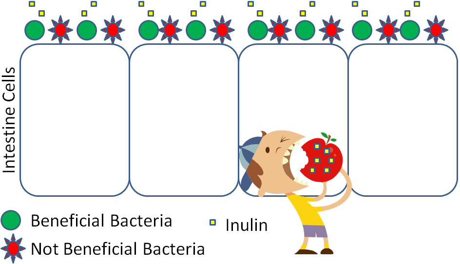
The net result is the same for both prebiotics and probiotics, an increase in the benign/not beneficial microorganism ratio.
The following video does a nice job of explaining and illustrating how probiotics work. The NCCAM website is a good source of information if you accept further questions on the topic.
Some common examples of probiotics are DanActive® and Activia®.
The claims that companies fabricated nigh their produce probiotic products have come under scrutiny. Dannon settled with the U.s.a. Federal Trade Commission to drop claims that its probiotic products will assist prevent colds or alleviate digestive problems, as seen in the top link below. General Mills also settled a lawsuit that accused them of a falsely advertising the digestive benefits of Yo-Plus a production it no longer sells, as seen in the second link.
Some examples of prebiotics include inulin, other fructose-containing oligosaccharides and polysaccharides, and resistant starch. Inulin is a polysaccharide that contains mainly fructoses that are joined by beta-bonds, which allows them to survive digestion. The structure of inulin is shown below.
Resistant starch is so named because information technology is a starch that is resistant to digestion. As a result, it arrives in the colon to exist fermented.
References & Links
- http://en.wikipedia.org/wiki/File:Inulin_strukturformel.png
- Douglas L, Sanders M. (2008) Probiotics and prebiotics in dietetics exercise. American Dietetic Clan.Journal of the American Dietetic Clan 108(3): 510.
Links
NCCAM: Probiotics – http://nccam.nih.gov/wellness/probiotics/
DanActive® – http://www.danactive.com/
Activia® – http://www.activia.us.com/
Danimals® – http://world wide web.danimals.com/New Campaign Markets Activia to Wider Audience – http://www.nytimes.com/2014/01/06/business/media/new-entrada-markets-activia-to-wider-audience.html?_r=0
General Mills Settles Yo-Plus Lawsuit – http://www.foodbusinessnews.net/articles/news_home/Site_News/2013/02/General_Mills_settles_Yo-Plus.aspx?ID={40F62478-1AA4-49DF-9330-E41E19E946D0}&cck=1
Video
Probiotics – http://www.youtube.com/watch?5=2k8Puxz54FQ&NR=i
Source: https://open.oregonstate.education/humannutrition/chapter/macronutrient-digestion/
Posted by: alvaresbeelty.blogspot.com
0 Response to "What Do Proteins Look Like Protein Digestion In The Stomach Animated Gif"
Post a Comment